In my earlier pages on poppycock terminology, I discussed the concept of
image magnification, and how it has been, intentionally or by
sheer ignorance, twisted and obfuscated in promotional and technical literature. Here I discuss the
magnification claims of a microscope of historical interest claiming to magically achieve magnifications
not allowed by photon physics, as well as some toy microscopes and webcam-based digital microscopes.
The realm of the possible
The resolution limit of conventional optical microscopes was established by Ernst Abbe around one and a
half centuries ago. Known as the
Abbe criterion, it
says that the smallest features an optical microscope can resolve are approximately half the size of the
wavelength of light. By using shorter wavelengths, one can resolve smaller features, but the shortest
wavelengths perceived by human eyes (around 400 nm) place a practical resolution limit at 200 nm for
visual observation.
Nothing prevents us to increase the magnification of an optical microscope beyond Abbe's limit, but this
only creates empty magnification that does not show any additional detail. For example, you could use the
eyepiece of a microscope to project an image on a large projection screen. The image will be very dark and
quite blurry, but it does work.
It can also be noted that, with a good optical microscope, it is entirely possible to detect the
presence of a particle of a diameter quite a bit smaller than the resolution limit of the
microscope, as long as individual particles are located conveniently far from each other. The particle,
however, is only visible as a featureless blurred circle, and multiple particles tightly packed together
are rendered as a single blur.
Additionally, Abbe's limit is the top theoretical limit to microscope resolution, but there is no bottom
limit to the resolution of a poorly built microscope. A good microscope for transmitted illumination with
air objectives and air condenser can reach 600x (typically with 10x eyepieces and 60x objective) and still
provide a visually sharp image, or 1,000x with a moderately fuzzy image. A better image quality at 1,000x
requires a 100x immersion objective and an immersion condenser. To substantially exceed this magnification
(e.g. by a factor of 100 or more), you need an electron microscope, X-ray microscope, or atomic-force
microscope.
We now know that it is possible to make smaller features than Abbe's limit visible with an optical
microscope by cleverly exploiting fluorescence and quantum phenomena. However, they require sophisticated
equipment like high-power lasers, near-field meta-optics placed at distances from the subject much smaller
than the wavelength of light, or
quantum-enhanced microscopes
that project quantum-entangled photons outside the range of near fields. You cannot find this level of
technology in a toy microscope (not for the next several decades, anyway).
Magnification of a microscope
Magnification of a microscope is specified as the ratio of linear image size seen through the eyepiece(s)
to linear subject size. The image size is measured on an image projected in front of the viewers' eyes at
the conventional distance of 25 cm.
In practice, magnification of a microscope is calculated by multiplying the magnification of the objective
by the magnification of one eyepiece, sometimes additionally multiplied by a tube magnification factor
specified on the microscope tube (typically between 1.25 and 2).
Rife microscope
Royal Raymond Rife, 1929, with one of his microscopes. By unknown author - Generally distributed on
Internet, CC BY-SA 4.0,
Wikipedia
The claims to fame of "inventor"
Royal Raymond Rife (1888-1971) are
an "oscillating beam ray" device said to be capable of curing almost any disease, and a microscope capable
of magnification far beyond any other optical microscope, in spite of using ordinary, commercially
available objectives and eyepieces. Some newspapers of the time, for example, reported that the Rife
microscopes were capable of a magnification of 17,000x. Other sources report claims of
30,000x.
The ray machine was likewise reported in the press to have cured several cases of terminal cancer within
60 days of treatment.
The numerous educational and honorary titles often attributed to Rife and his purported early experience
as a Zeiss microscope technician have remained unsubstantiated, despite investigations. His documented
work experience is as a private driver and car mechanic for a well-to-do man.
A search on
Quackwatch returns
two pages of results about Rife and his followers, several of whom have been convicted of fraud (mainly
for selling "medical" devices not approved and not effective, and for promising to cure cancer
with "alternative" medicine). If you really feel like it and have no better use for your time,
you can read a recently published
313-page PDF
written by Rife's followers, describing Rife's accomplishments, devices and theories.
Aside for an internal prism system designed to increase the length of the optical path inside the
microscope tube, the Rife microscopes available for inspection and/or documented in photographs do not
appear to contain anything capable of fulfilling Rife's claims. In a finite microscope, increasing the
optical tube length does increase magnification, but does nothing to increase image resolution. Usually,
it decreases the latter by forcing the objective to work outside of its design parameters. Naturally, some
proponents of Rife microscopes maintain that a "secret" component has been removed from these
microscopes, and even wrote lengthy "explanations" involving multiple stacked optical slits and
other unworkable devices. Unfortunately, all these discussions are purely qualitative, and ignore basic
aspects of light propagation and diffraction.
There are indeed ways to go slightly beyond Abbe's limit even without quantum-enhanced optics, e.g. by
using a slit-like diaphragm instead of a round one. Indeed, some reputable makers of research microscopes
used the same method in some of their special-purpose microscopes, where measuring the subject in a single
direction was desirable (e.g., the thickness of a fiber, while the length of the fiber and its internal
texture were irrelevant). However, this slightly increases resolution in one direction, while
simultaneously reducing it in a perpendicular direction. Stacking two slit diaphragms at a right angle to
each other, as some of the "explanations" of the Rife microscope propose, produces no net
increase in resolution, only a net loss of resolution caused by diffraction in both slits.
Other explanations are simpler nonsense, like the assertion that to make the Rife microscope reach the
advertised magnification it is necessary to replace the eyepieces with objectives. This conveniently
ignores "details" like the different location of entrance/exit pupils in objectives vs.
eyepieces.
Bracegirdle (2003)
provided an account of the Rife microscopes, including one he was able to inspect and test. Like numerous
other scientists and microscope experts, he concluded that Rife's claims are impossible. He further
suggested, and I concur, that the extremely high number of unnecessary fine mechanical adjustments of the
Rife microscopes appears designed to impress laymen, but make the use of the microscope so fiddly to
become, at best, impractical. Multiple accounts, for example, report that it took hours of fiddling before
being able to obtain an image from a Rife microscope (even by specially trained Rife technicians), most
often of such a poor quality as to be useless. Additionally, a few faults and oversights in its design
betray the inexperience of the designer. I believe the unnecessary mechanical complexity is partly smoke
and mirrors, and partly a subterfuge to deflect criticism that the device does not work by counter-arguing
that the user failed to properly "calibrate" it.
At one point, Rife sold a microscope and a beam ray device to a European company interested in importing
and marketing these devices, and even sent a trained technician along to set up the microscope. This
company was never able to obtain any of the claimed results, the microscope did not work despite the
on-site attempts by the technician, and in the end Rife simply stopped replying to their inquiries.
None of the web sites reported at the end of Bracegirdle's paper are still active, although I accessed at
least two of them a few decades ago and was able to find them again on the
Wayback Machine.
Rife's claims about extremely small "microbes" causing all diseases, and being able to morph
into each other, as well as to and from a multitude of different shapes and sizes, are known as Rife's
pleomorphism "theory". An additional claim by Rife is that each of these microbes can be
vanquished by exposing a patient to a specific radio frequency, or set of frequencies. These claims are
frequently repeated and amplified on short-lived web sites promoting "alternative" views on
science and medicine, especially cures for cancer. Some of these web sites also publish discussions of the
Rife microscopes, probably to reinforce the credibility of the claims about these "microbes" not
visible with any other instrument than Rife microscopes, and indirectly of the claims about the effects of
"oscillating beam ray" machines.
Later in his life Rife himself, perhaps fearful of being sued for false medical claims, had become
slightly more careful with his claims, and limited himself to stating that his microscopes, rather than
providing a so-far unheard-of high magnification (which is a demonstrable falsehood even without testing
his microscopes), were particularly good for "special applications". This included observing the
"microbes" he claimed caused most or all diseases and could be treated by his other main
invention, the "oscillating beam ray". Needless to say, no one else could find these
shape-shifting microbes with the best medical microscopes of the time, nor with the electron microscopes
developed shortly thereafter, and there is no credible documentation of the beam ray devices successfully
treating any diseases. Some beam ray devices were tested and found to be DOA, with disconnected internal
wiring - they simply did not turn on, nor did anything at all.
Some modern variants do work, in the sense that they produce a visible electrical discharge inside a
gas-filled tube and a buzzing sound, but so does a neon sign, and there is no medical proof that neon
signs and fluorescent tubes cure diseases like terminal cancer and advanced-stage viral infections. None
of the oscillating beam ray devices nor their modern varieties actually emits the frequencies they are
supposed to, described in Rife's own descriptions and lists.
The American Medical Association eventually forbade Rife from treating patients with his machine, and he
died penniless in a hospice.
Barry Lines is the author of at least five books promoting Rife's cancer "cure", still available
on Amazon:
1,
2,
3,
4,
5. A search for "cure for cancer" on Amazon returns over 9,000 hits. Hundreds of other books,
booklets, web pages and other materials of a similar character can be found on the web, as part of a
veritable rabbit hole to an alternate universe of "vibrational medicine",
"electromedicine", "holistic health", "electron therapy", "orgone
accumulators", "pyramid power", "magnet therapy", "phrenology",
"radionics" etc., along with materials of historical interest including e.g.
radioactive quackery
promoting the "therapeutic" effects of ingesting radium, thorium and other radioactive materials
(which did directly cause the well-publicized
death of an industrialist and quite
possibly other deaths and diseases).
Although several of these materials have been officially unmasked as quackery (here is an example of
original claims
and
their debunking), many more keep popping up like mushrooms after an autumn rain. The 1940
electro-metabograph
by the Art Tool & Die Company of Detroit, for example, is much flashier that Rife's sparingly-built
oscillating beam ray device, although based on similar (false) principles.
The ADE 651 and HEDD1 bomb detectors
An interesting conceptual variant of the "oscillating beam ray" scam emerged after the
Coalition-Iraq wars, when the Iraqi government was scammed by the British company ATSC into buying very
expensive remote detectors of bombs
and mines capable of detecting explosive devices at distances up to kilometers (and even drugs, ivory, and
other chemicals and materials).
This scam is related to Rife's oscillating beam ray machine for using "the interference between the
magnetic field of the earth, the explosive, the device itself, and the human body" (quoted from the
company's description of how its detectors work). The
financial part
of the scam is slightly more complicated than Rife's, and involves using false public claims that the
device works in order to artificially inflate the company's stock prices, then selling the stock to
unsuspecting investors before it crashes.
The original Sniffex and the subsequent Sniffex Plus were, essentially, empty plastic boxes equipped with
a handle, a few fake controls, and a
dowsing rod.
Not even a battery. After a while, a company called Unival did solve the difficult technological problem and started
selling the
HEDD1
device. This revolutionary new device does have a battery compartment (batteries not included).
This article
provided by Sniffex Plus and HEDD1 in 2010 describes both the original battery-free device and the
improved device with battery as a resounding success (the websites for both companies are now dead).
Incidentally, there was a different SniffEX device from a different company that worked, and is not a
scam. It was originally called Sniffex but the name was not trademarked, so ATSC successfully sued the
original Sniffex makers and forced them to change their device name.
The ATSC detectors were configured by plugging into the device a cartridge that had been
"activated" by storing it for some time together with a sample of the substance to be detected.
The makers of these "detectors", confronted with accusations that the devices were a simple
fraud, claimed that the person holding the box, and the mental attitudes of this individual, were
essential components of the detector. This was tantamount to saying that the detector only worked if held
by a person firmly believing that it did work, and any skeptic wishing to test the device would cause it
to fail. Any detector user blown up by an undetected bomb died solely because of his/her own fault.
At least three other commercial devices (one of them originally sold to locate lost golf balls) are very
similar to the ADE 651 and its variants. Together with the latter, all these devices were eventually
banned from use by military and security in multiple countries, and several of the persons involved in
their marketing jailed because of the stock fraud associated with the scam. Apparently you can go to jail
and/or be forced to pay millions of USD for stock market fraud and for selling security equipment that
does not work to military and police, but rarely for selling cancer cures that don't work to sick
individuals.
A modern Rife microscope
For the past 48 years, a company called
has been running a web page announcing the impending release of what appears to be a modern Rife
microscope, with similar claims to the latter and likewise using ordinary commercial objectives and
eyepieces. Like the Rife microscopes, they are characterized by extreme mechanical complexity, some of it
evidently purposeless even for observers with only a moderate experience of microscopes.
The only developments reported by this company in these 48 years are:
The death, in 2022, of the "German microscope expert" involved in this project.
A promise to start delivering microscopes in "Q3, 2022", and a text blaming the Covid pandemic
for delays.
A "nominal" charge of 400 USD for demonstrations of their microscope in Hamburg, Germany.
The company is apparently run from a Las Vegas P.O.Box, with labs and personnel located "in
Germany". I will not link to their web site, but if you really want you can easily Google it. Google
will also give you an address for the company in Cologne, Germany, which turns out to be a residential
apartment block.
The Scientific Papers tab of the company's web site begins with a promising paragraph:
"A number of scientific papers have been written using the results obtained with" the company's
microscopes, and continues: "We are offering some of these papers for download without further
comment." The only paper, scientific or otherwise, available for download is a three-part PDF in
German, entitled "DerBlickindieUnendlichkeit". It consists mostly of a haphazard
discussion of Rife's pleomorphism "theory" and other pseudo-scientific babble, together with
some claims about the company's microscopes.
As of March, 2023, the Scientific Papers tab on the US web site (.com top domain) is
still available, albeit not on the German (.de) web site. It is also available in 92
snapshots of the company's US web site on
Wayback Machine. The PDF is also available on
Yumpu, together with several other tell-tale documents from the same microscope company (e.g.
1,
2,
3,
4,
5,
6). I regard it as highly significant that a text in German was made available on the US web site of the
company, where most visitors would not be able to read it, but not on the company's German web site, where
any local visitor with a minimum of technical or scientific education would be able to read it and debunk
its claims.
The PDF is illustrated with pictures reportedly taken by the deceased microscope expert with one of the
company's microscopes.
The only significant bits of information worth discussing that I found in this PDF:
The assertion, in Part 3, Figure 7, that one of the company's microscopes is capable of a 100 nm
resolution, while "ordinary light microscopes" only reach a 400 nm resolution. The purpose of
the figure is to assert that the company's microscopes have four times the resolution of an ordinary
optical microscope. However:
A 100 nm resolution is available by using a 200 nm UV light source and good UV optics (both are
commercially available from multiple sources). This capability is far from unique for an optical
microscope.
The assertion that ordinary light microscopes stop at a resolution of 400 nm is wrong. 400 nm is
violet light still within the visible range, and the resolution at this wavelength is 200 nm according
to Abbe's criterion. Therefore, any good visible-light microscope can reach the resolution of 200 nm
by using a monochromatic 400 nm illumination, without special optics and still allowing visual
observation through the microscope. UV microscopes with a resolution of 170 nm are not too difficult
to assemble from off-the-shelf parts that have been available for decades, and obviously require
digital cameras or other devices to convert UV to visible images.
The assertion in Part 3, Figure 8 that the company's microscope provides (1) a resolution of 100 nm, (2)
an enormously higher DOF than other optical microscopes, (3) a
"magnification up to approx25,000x true-color images". This can only prompt one reaction: Poppycock alert! Breaking down the statement:
The 100 nm resolution claim is possible, as already discussed, and far from unique.
An unlimited DOF can be obtained, with static subjects, by using an image fusion technique called
focus stacking, popular in
photomacrography, also common in
optical microscopy, available in many digital cameras, and even used on Mars by the
Curiosity rover. It is
not a unique capability of the microscopes of this company. It can be used with any conventional
optical microscope equipped with fine focus, after a mechanical modification of this focuser. For
example, I am using it on a 35 year old Olympus compound microscope, and some microscopists use it on
microscopes built a good one century ago.
The 25,000x magnification, on the other hand, is pure, unadulterated poppycock (unless it is empty
magnification that shows no more detail than at 1,000x - but what good does it do?). You need a good
SEM, TEM or STEM to get 25,000x. As for the "real color" part (a claim also repeated several
times on the company's web site), at 25,000x there can be no color because we are at a scale far
smaller than the wavelengths of visible light. Hollywood can easily show you the quantum world in
dazzling colors, but physics and technology have their limits. Even at 200 nm illumination (100 nm
resolution) there can be no "true color". Color is at wavelengths between 400 and 700 nm.
Like the company's web site, the text of this PDF seems to have been gradually sanitized of some of the
most obvious false claims relating to microscopy, but the figures and their captions seem to have been
re-used from earlier materials and have not been revised. The real purpose of the PDF is promoting one of
the many variations on the theme of Rife's pleomorphism, especially for what concerns cancer and its
"alternative treatments". It would be easy to laugh these claims off, were it not that they are
designed to entrap gravely sick people hoping to find a cure, or their desperate relatives grasping at
straws to find help for a terminally sick family member that physicians no longer can help, and
financially exploit their suffering and trust for financial gain, in return for nothing more than false
treatments, false promises and false hopes. There are also several documented cases of patients quitting
medical treatments that could potentially have saved or extended their lives in order to follow
quackery advice.
There isn't much money to be made by selling microscopes that don't work and have never been available for
purchase, so I am convinced that this microscope company is only a side show to increase the credibility
of the main scam of selling miraculous treatments that
have no basis in science.
I failed to find any information on the supposed journal on which the PDF was published, let alone whether
it really was published. Publication on a formally established and indexed science journal, in itself, is
no proof of scientific value, because there are thousands of indexed "scientific"
predatory journals with
high sounding names (sometimes highjacked from respectable journals of the past that are no longer
published, other times similar enough to respectable journals to be confused with the latter) that will
publish virtually anything as long as the author pays a substantial charge. These journals are mostly
based in India, where a single predatory publisher may own a roster of several hundred journals on the
most disparate subjects, but quite a few operate from other countries. I myself regularly receive
invitations to submit papers for publication in such journals and/or to participate in their editorial and
review boards. Quite a few respectable scientists have been duped into lending their prestige and
professional credibility to these journals, at least when predatory publishing was still a relatively new
phenomenon.
The name of the author of this PDF is found on the web in connection with a few privately published
booklets and texts expounding the same general type of pleomorphism theories and other unproven poppycock.
One example
talks about "blood flow paintings" that have no place in real medical science, at least in our
universe, and
another
on isopathy, which is either a variant of, or a a less common name for,
homeopathy. The latter is an "alternative" method of treating diseases popular in Germany, but based on
premises not supported by medical research. These booklets are apparently designed to attract the
attention of cancer patients and other people suffering from medical conditions that medicine cannot yet
tackle successfully. The author is also listed as a "health practitioner" in a small German
town.
For reasons I don't fully understand, even a few modern countries like Germany have a long tradition of
allowing "parallel" private medical businesses and practitioners with resounding but
scientifically questionable titles and educational degrees to apply "alternative" treatments of
doubtful or no medical value on paying private patients.
The Scientific Papers tab on the company's web site additionally links to a few
promotional videos, but nothing that could qualify as a scientific paper.
I will not further discuss Rife's claims or those of his followers, other than reporting that the
transquantum-based Bull field detector in my lab, which I carefully built after many years of secret
research, gives off-the-scale readings when brought close to pictures and descriptions of Rife's and his
followers' microscopes and beam ray devices.
Disclaimer: The plans for building my Bull field detector are secret and not for sale
(yet - but feel free to ask me again in another 48 years).
Kang & Pedersen (2017) devoted less than a page of their book on quackery to Rife and his
inventions.
Reference:
Kang & Pedersen, 2017: Quackery, a brief history of the worst ways to cure everything. 492 pp, Workman
Publishing Company.
Magnification of toy microscopes and digital microscopes
Some cheap toy microscopes are, in a way, comparable to Rife microscopes in the sense that their
promotional materials describe them as capable of absurdly high magnifications.
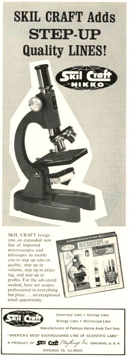
Vintage ads for a low-end microscope and a toy microscope.
These particular ads do not make exaggerated magnification claims.
In the past, exaggerated magnification claims were especially common in ads for toy microscopes on the
back cover of comic books. A common trick in these ads was of using a "non-standard" definition
of magnification: instead of using linear magnification, they published a figure computed as area
magnification, usually without mentioning it, or only as a footnote. In this way, a microscope with 10x
eyepiece and 10x objective (100x in total) suddenly became capable of 10,000x magnification (100 times
100). One with 10x eyepiece and 50x objective became a 250,000x microscope, and so on.
These magazine ads have largely disappeared, together with those for "spy cameras" one could
hide in the palm of one's hand and "X-ray sunglasses" capable of penetrating women's clothes to
reveal their underwear. I still see ads for toy microscopes reaching 1,200x, which is not outside the
realm of the possible (especially if it is empty magnification). However, I see a possibly misleading way
to specify the magnification of digital microscopes based on cheap webcams, typically advertised on Taobao
and Aliexpress, and now making their way also onto Amazon. For example, the following ad titles are common
on Aliexpress. Ads and seller names change frequently, so perform a search on Aliexpress.
Portable 600x 4.3" LCD Digital Microscope 8 LED 3.6MP VGA Electronic HD Video Microscope PCB
Repair Endoscope Camera Magnifier
1200X Microscope Digital Portable 7" LCD Video Microscope 12MP for Soldering Electronic PCB
Inspection Continuous Zoom
AliExpress ad for a "600x digital microscope". Note the lack of a focusing mechanism.
Since these microscopes are designed for repairing electronic PC boards, the 1,200x type has only coarse
focusing, and the 600x type often entirely lacks a focusing mechanism. You focus it by moving the subject
held in your hands, or by bending a scarcely practical plastic gooseneck with plenty of backlash. A 600x
or 1,200x real magnification, measured as customary for microscopes, would make focusing by these means
impossible. Such a magnification would also be way too much for the job of repairing PC boards, and would
require sophisticated and expensive optics to provide the working distance of several cm shown in the ads.
The NA of these long-working-distance optics would necessarily be low, given the small diameter of the
objective.
The magnification specified in the ads is also suspiciously close to that obtained by squaring the linear
magnification estimated for these microscopes (252 = 625, and 352 = 1,225), so I
cannot exclude that these ads refer to area magnification instead of linear magnification. They may be, in
this respect, a reincarnation under a new guise of the misleading magazine ads for toy microscopes.
A 300 Mpixel microscope camera for a few bucks?
eBay ad picture and text for a "300 Mpixel" microscope camera module.
At present, dozens of China-based eBay sellers advertise a
300 Megapixel Microscope Module Electronic Eyepiece Camera Module USB2.0 output for around 12-15
USD. A 300 Mpixel digital camera to mount on a microscope, if you can find such a thing, should cost
thousands of USD. Most likely you cannot find it, because 300 Mpixel would be a terrible waste of image
resolution, considering that 30 Mpixel is probably already overkill for a conventional optical microscope.
Additionally, a 300 Mpixel sensor is wasted unless the lens is capable of a comparable resolution, and I
can tell you that a 300 Mpixel lens would cost more than a good research-quality microscope.
I cannot imagine a cheap plastic lens with a front optical element 10 mm in diameter having such an image
resolution, let alone a 300 Mpixel sensor fitting in the space behind this lens.
Additionally, a USB 2.0 interface as described in the ad would take minutes to transfer a single 300
Mpixel image (almost 1 GB uncompressed for a 24 bit-per-pixel color image).
To put things in perspective, an 8k camera sensor or LCD screen has a resolution of around 33 Mpixel, and
the today still rare 16k is about 132 MPixel. These ads are just an example of the current
Ferengi-like attitude of many Chinese
retailers that "anything goes, as long as I make a profit".
From the price and picture, I guess they are offering, in the best case, an old and outdated 3 Mpixel
generic webcam module (the picture shows a module with a readable 2015-05-19 date on the PC board). You
can even see where a number of small LEDs are supposed to be soldered to the PCB around the lens mount, so
obviously this module was designed as a simple security camera or webcam. A proper microscope camera, i.e.
a camera designed to be mounted on a microscope, has no use for LEDs around the lens. I can see no way a
honest mistake could cause the resolution to become advertised as 300 Mpixel.
Conclusions
The Rife microscopes are an outstanding example of pseudo-scientific fraud, designed
mainly to support and lend credibility to other pseudo-scientific claims of
curing/treating a broad range of diseases with an "oscillating beam ray" machine of no
medical value. More modest examples of misleading magnification claims can be found in
old advertising of toy microscopes and current webcam-based "digital microscopes". Current eBay ads by multiple Chinese sellers
misrepresent a cheap and outdated webcam module as a 300 Mpixel microscope camera.